Blue light is a part of visible light (visible to the naked eye as blue); the wavelength of about 400 ~ 500nm, it is a high-energy short-wave blue light. It is not only seen in daylight, and a large number of LED lamps, neon lamps, mobile phones, computers, and other electronic devices. Studies have shown that the energy of blue light is weaker than ultraviolet light, but can directly penetrate the dermis layer of the skin to cause direct damage to the skin, so anti blue light compositions for skin care is necessary. Blue light irradiation induces the onset of skin ageing by inducing ROS production, leading to a decrease in type I collagen synthesis and an increase in MMP1 synthesis in skin cells, which further degrades collagen.

Figure 1. Different wavelength
Harmful effects of blue light on the skin
- Induction of skin pigmentation
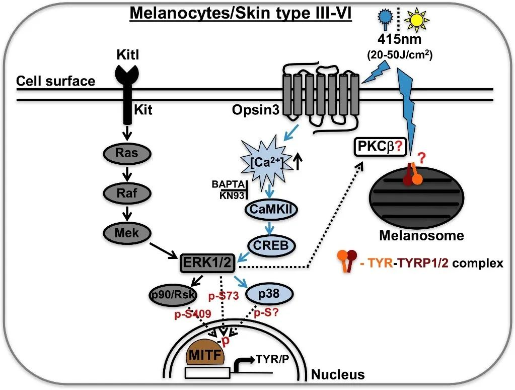
Melanocytes can directly perceive blue light in visible light at a wavelength of 400-415nm through OPN3 (Opsin-3), causing it to generate a protein complex of tyrosinase and dopa pigment interconverting enzyme, which continuously activates tyrosinase and leads to skin pigmentation. That can result in skin to become discoloured, dull, loss of luster, uneven skin tone and other problems.

Figure 2. Blue light causes skin pigmentation
- Accelerated skin photoaging
Photoaging is skin aging caused by sunlight exposure. Exposure to certain wavelengths of blue light causes oxidative stress and cytotoxicity in cells and reduces the antioxidant capacity of fibroblasts; it also inhibits fibroblast proliferation, and reduces the extracellular matrix, leading to photoaging of the skin. Skin problems such as hyperpigmentation, skin inflammation and wrinkles will follow.
- Disruption of physiological rhythms
Skin cells are very sensitive to blue light, which directly affects the production of free radicals. In other words, blue light disrupts the body’s circadian rhythm and accelerates skin aging. That’s why playing with your mobile phone before bed is more likely to make you feel that your skin is in poor condition.
There is a study which showed the compositions of hyaluronic acid and ectoin have the anti blue light effect.
Hyaluronic acid in anti blue light compositions
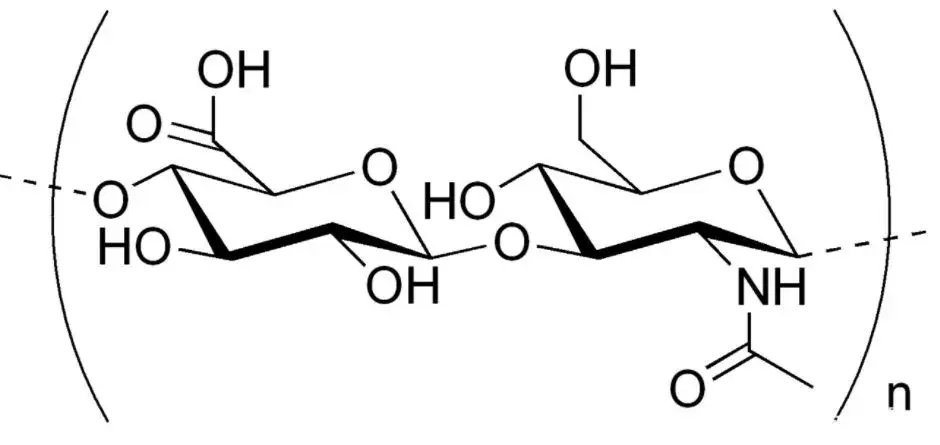
Hyaluronic acid (HA) is a straight-chain polymer polysaccharide with disaccharide (a disaccharide derivative consisting of N-acetyl-D-glucosamine and β-D-glucuronic acid linked to disaccharide derivatives by β-1,3-glycosidic bonds) as the repeating unit, and its structural formula is shown in below figure. In 1934, Karl Meyer and John Palmer of Columbia University in the United States isolated HA from the vitreous body of the cow’s eye and named it “Hyaluronic Acid”, which is derived from the words “Hyaloid” and “Uronic Acid”. Later, in 1986, Endre Balazs coined the term “hyaluronan” to name hyaluronic acid in order to comply with the international nomenclature of polysaccharides, which covers a wide range of molecular forms (both acid and salt forms).

Figure 3. Structural formula for hyaluronic acid
Ectoin in anti blue light compositions
Ectoin is an amino acid derivative with powerful anti-stress protection that protects extremophilic microorganisms and plants against lethal, extreme elements of the environment in which they live, such as high salinity (salt lakes), severe cold (glaciers), and dryness (deserts). Ectoin has a strong ability to complex with water molecules, forming a “hydration shell” around cells and proteins, effectively locking the water and preventing the loss of skin moisture, thus achieving the moisturizing effect. Secondly, ectoin can stabilize the cell membrane and protect the cells from external environmental damage, such as UV radiation, pollutants, heat and cold, etc., to enhance the skin’s resistance. In addition, ectoin can promote collagen synthesis, enhance skin elasticity, reduce fine lines and wrinkles, and slow down skin aging.

Figure 4. Structural formula for ectoin
The anti blue light compositions of 1.5% hyaluronic acid, 4.5% ectoin and 0.1% ergothioneine, dissolved with water and stirred until homogeneous.
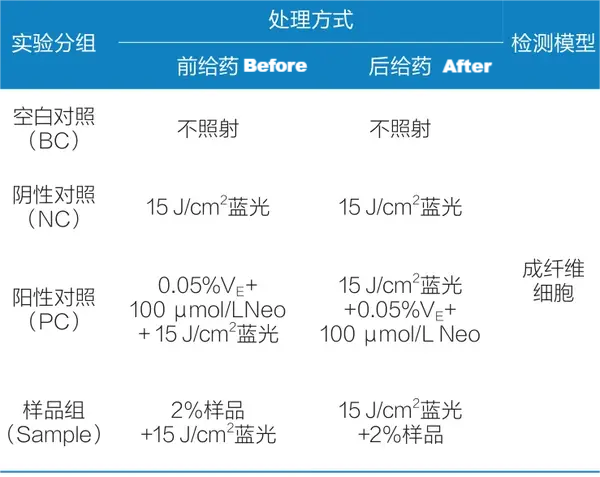
The experimental protocol is shown in figure 5, 2 mL of cell culture medium was added to each well of the blank control group and negative control group; 2 mL of culture medium containing 0.05% VE and 100 μmol/L Neo was added to each well of the positive control group; 2 mL of culture medium containing the corresponding concentration of the test material was added to each well of the sample group (2% anti-blue-light compositions); and the negative control group, the positive control group, and the sample group were subjected to blue-light irradiation at a total dose of 15 J/cm² of blue light irradiation, meanwhile, the blank control group was placed in the same environment but without blue light irradiation.

Figure 5. Test scheme of anti-blue light effect
As shown in Figure 6, compared with the negative control group, the sample group (2% anti-blue light composition) significantly inhibited the elevation of reactive oxygen species (ROS) induced by blue-light irradiation in both before and after 2 modes of administration, with an inhibition rate of 9.99% in the before, and an inhibition rate of 17.55% in the after. This suggests that the anti-blue light compositions can not only inhibit the elevation of reactive oxygen species (ROS) induced by blue light irradiation, but also scavenge the oxygen radicals generated under blue light irradiation.

Figure 6. Trends in ROS content before and after
As shown in Figure 7, in the pre-administration mode, the sample group (2% anti-blue light combination) significantly promoted the secretion of Collagen I with an enhancement rate of 41.28%; and in the post-administration mode, the sample group (2% anti-blue light combination) significantly promoted the secretion of Collagen I with an enhancement rate of 113.02%.

Figure 7. Trends in Collagen I content before and after
As shown in Fig. 8, there was no significant change in the MPP-1 content of the sample group (2% anti-blue light combination) in the pre-administration mode compared to the negative control group. Whereas, under the post-dosing mode, the MPP-1 content of the sample group was significantly reduced with an inhibition rate of 36.33%. It indicates that the composition can inhibit the increase of MMP-1 content caused by blue light irradiation, and thus has the effect of inhibiting collagen decomposition.

Figure 8. Trends in MMP-1 content before and after
The 2% concentration of the anti-blue light composition (1.5% hyaluronic acid, 4.5% ectoin and 0.1% ergothioneine) can effectively inhibit the elevation of reactive oxygen species (ROS) induced by blue light irradiation, promote the secretion of Collagen I, and prevent the damage of blue light. Especially after blue light irradiation, the product can also increase the activity of intracellular SOD enzyme and inhibit the expression of MMP-1 to improve the photo-ageing caused by the damage of blue light. The product can also increase the activity of intracellular SOD enzymes and inhibit the expression of MMP-1 to improve the problem of photoaging caused by blue light damage.
Related reading
- Ectoin introduction
- Ectoine Uses In Skincare Products
- Ectoin Skin benefits for anti-inflammation & skin repair
- Hyaluronic Acid Benefits for Skin Care
- Hyaluronic Acid Moisturizing Effect In Cosmetics
- Ergothioneine Introduction


